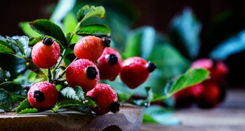
几个世纪以来,玫瑰果一直是草药的主要原料…
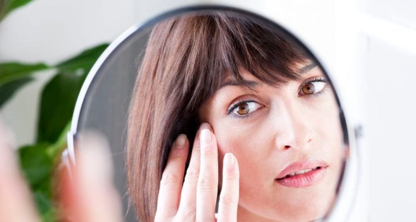
随着岁月的流逝,皮肤会经历自然老化的过程…
尿失禁是一种常见、令人尴尬和不快的健康问…
减肥是一个多方面的复杂过程,由几个同等重…
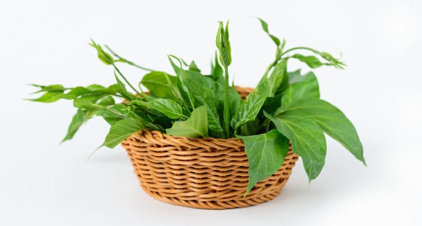
Gurmar 是一种源自亚洲的药用植物,…
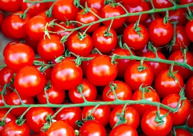
番茄红素是最强大的抗氧化剂之一,在我们体…
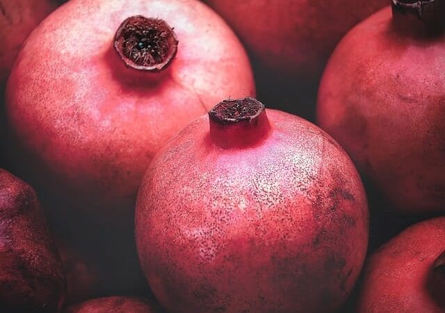
石榴原产于亚洲,是一种非常美味、特别多汁…
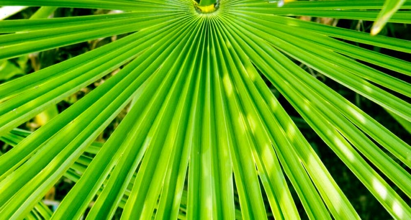
沙巴棕是一种北美药用植物,在解决良性前列…
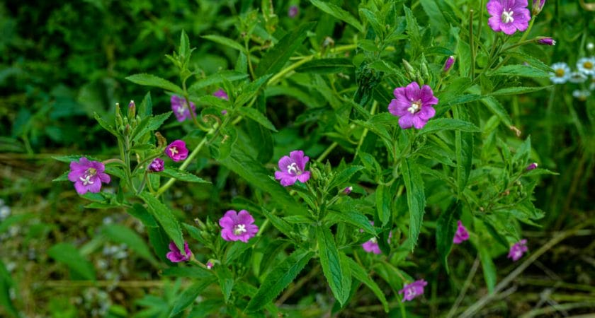
在 19 世纪,小花柳叶菜是治疗细菌性痢…