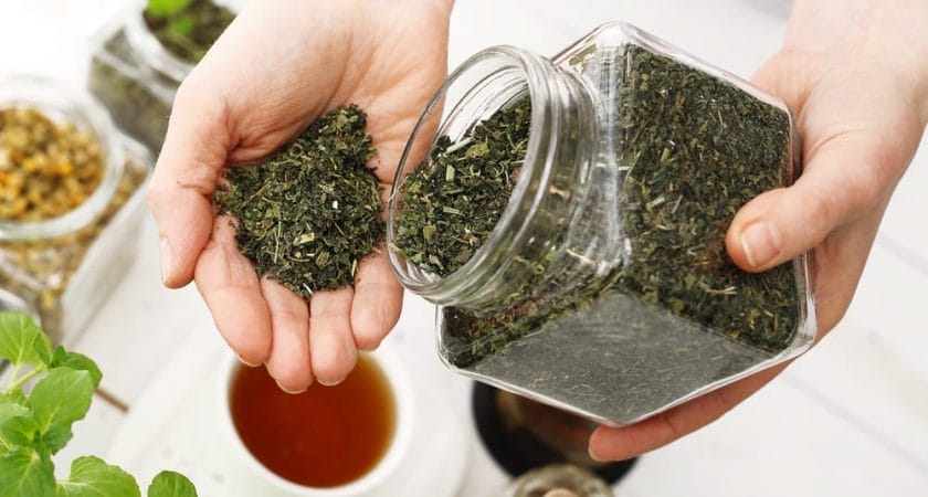
草药是一种非凡的健康盟友,从遥远的古代起…
普通感冒是秋冬季节的常见病。在防治感冒的…
无论男女,饮食和其他生活方式对生育都很重…
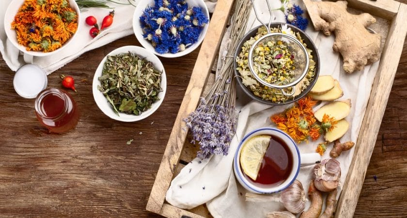
我们越来越多地在寻找提高免疫力和保持健康…
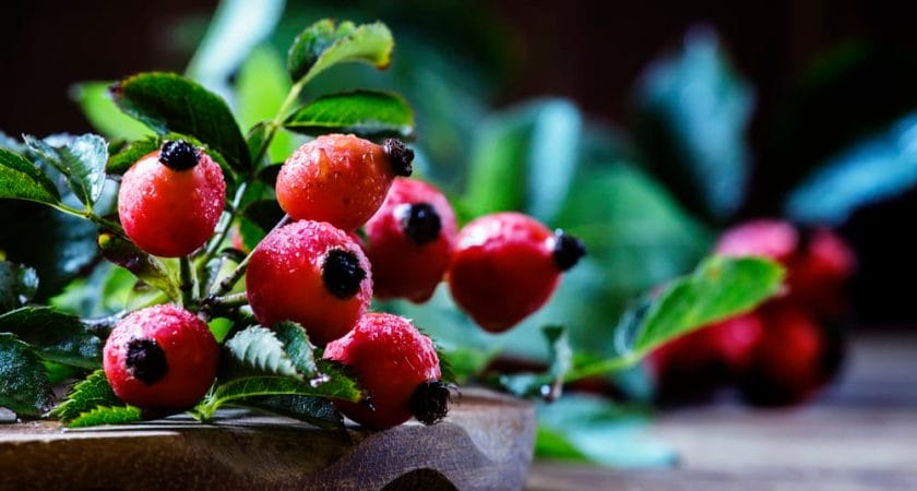
几个世纪以来,玫瑰果一直是草药的主要原料…
随着岁月的流逝,皮肤会经历自然老化的过程…
尿失禁是一种常见、令人尴尬和不快的健康问…
减肥是一个多方面的复杂过程,由几个同等重…
Gurmar 是一种源自亚洲的药用植物,…